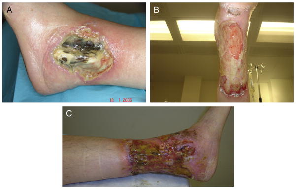
Fig. 1.

(A) Lateral malleolar ulcer at presentation demonstrating central necrosis of the tendons with extensive inflammatory slough and deep overhanging gunmetal gray borders consistent with pyoderma gangrenosum. (B) and (C) Progression of ulceration to become circumferential by 3 years after presentation.
