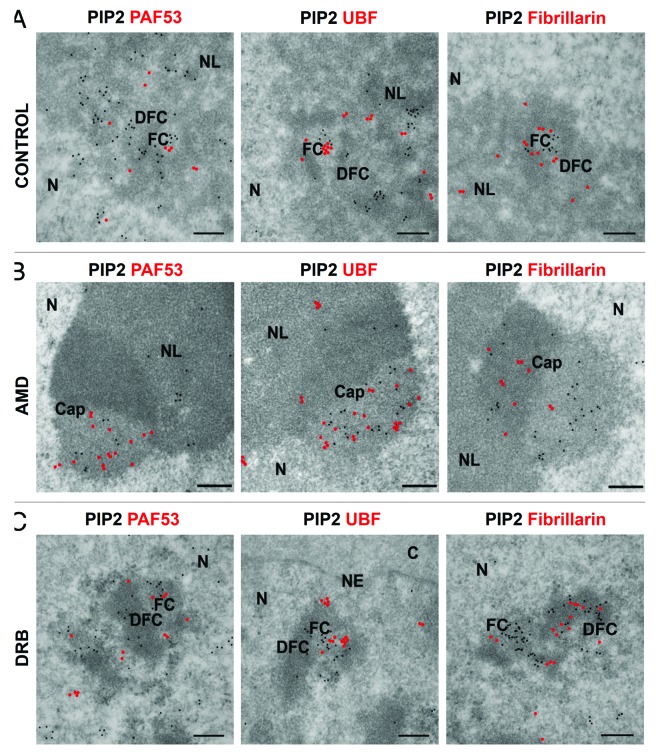
Figure 5. PIP2 colocalization with Pol I and UBF is not influenced by transcription inhibition while PIP2 colocalization with fibrillarin is disrupted by transcription inhibition as shown by IEM. (A) IEM results show that PIP2 is in close proximity to Pol I in the nucleolus and colocalizes with UBF in the FC and with fibrillarin in DFC regions, respectively. (B) In AMD inhibited cells, PIP2 localizes to the light part of the caps together with Pol I and UBF while fibrillarin localizes mainly to the denser part of the caps after the inhibition of Pol I transcription. (C) Upon DRB treatment, PIP2 colocalizes with Pol I and UBF in the inner space of FCs as well as on the border between FC and DFC regions, while in the DFC, PIP2 and fibrillarin are arranged in a necklace-like manner. N, nucleus; NL, nucleolus; FC, fibrillar center; DFC, dense fibrillar component. Scale bar: 200 nm.

An official website of the United States government
Here's how you know
Official websites use .gov
A
.gov website belongs to an official
government organization in the United States.
Secure .gov websites use HTTPS
A lock (
) or https:// means you've safely
connected to the .gov website. Share sensitive
information only on official, secure websites.
