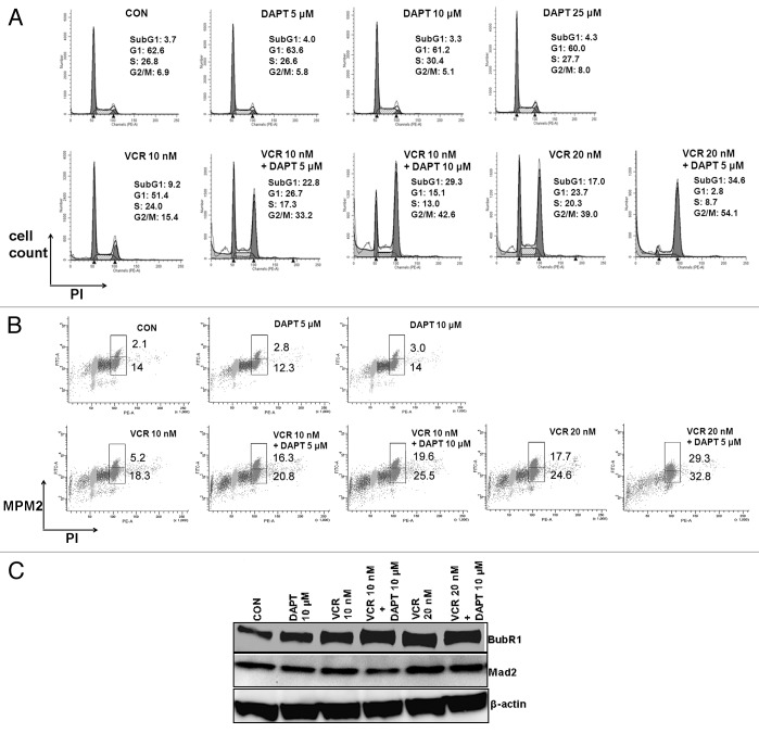
Figure 2. GSI augments VCR-induced mitotic arrest in a dose dependent manner. HeLa cells were treated with increasing concentrations of DAPT (5, 10, 25 μM) and/or VCR (10, 20 nM) for 24 h. (A) Cell cycle progression was analyzed after PI staining. The percentage of cells in each cell cycle phase is presented. (B) Cell population in mitotic phase was measured by double staining with PI and MPM-2. The percentage of cells in M phase (upper box) and G2 phase (lower box) are presented. (C) Cell lysates were analyzed for BubR1 and Mad2 by western blot. Beta-actin served as a loading control.

An official website of the United States government
Here's how you know
Official websites use .gov
A
.gov website belongs to an official
government organization in the United States.
Secure .gov websites use HTTPS
A lock (
) or https:// means you've safely
connected to the .gov website. Share sensitive
information only on official, secure websites.
