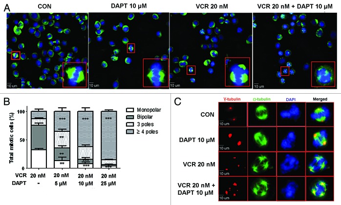
Figure 4. GSI amplifies VCR-triggered multi-polar mitotic spindle formation. HeLa cells were treated with VCR (20 nM) alone and/or increasing concentrations of DAPT (5, 10, 25 μM) for 24 h. (A) Representative examples of mitotic spindles stained with anti-α tubulin (green) and DAPI (blue) for the indicated treatment. Scale bars represent 10 µm. Inserts show representative image from each sample at higher magnification. (B) Observations demonstrated in (A) were quantified by evaluating at least 100 cells per treatment. Untreated control and DAPT treatment alone were left out from analysis because of lack of abnormality in mitotic spindles. Data from three independent experiments were graphed as a stacked mean percentage ± SEM. The statistical significance of differences was determined by ANOVA test. Statistical P values shown in figure were calculated with comparison to VCR treatment alone (*P < 0.05; **P < 0.01; ***P < 0.001). (C) Representative immunofluorescence images of mitotic spindles stained with anti-α tubulin (green), centrosomes stained with γ-tubulin (red), and DAPI (blue) for the indicated treatment.

An official website of the United States government
Here's how you know
Official websites use .gov
A
.gov website belongs to an official
government organization in the United States.
Secure .gov websites use HTTPS
A lock (
) or https:// means you've safely
connected to the .gov website. Share sensitive
information only on official, secure websites.
