Figure 5.
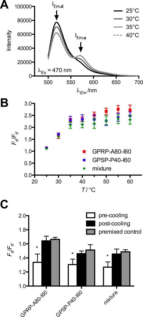
Formation of mixed micelles demonstrated using FRET. (A) Representative emission spectrum obtained from an equimolar mixture of GPRP-A80-I60-AF488 and GPSP-P40-I60-AF546 (1 μm final protein concentration) excited at 470 nm at various temperatures. Emission peaks of donor AF488 (IEm,d) and acceptor AF546 (IEm,a) are indicated by arrows. (B) Results are reported as fold change in IEm,a (Fa = IEm,a/I0Em,a) divided by fold change in IEm,d (Fd = IEm,d/I0Em,d) for equimolar mixtures of GPRP-A80-I60-AF488 and GPRP-A80-I60-AF546 (squares), GPSP-P40-I60-AF488 and GPSP-P40-I60-AF546 (circles), GPRP-A80-I60-AF488 and GPSP-P40-I60-AF546 (diamonds) as a function of temperature. (C) Fa/Fd for mixtures of pre-formed micelles of GPRP-A80-I60-AF488 and GPRP-A80-I60-AF546 (GPRP-A80-I60), GPSP-P40-I60-AF488 and GPSP-P40-I60-AF546 (GPSP-P40-I60), GPRP A80-I60-AF488 and GPSP-P40-I60-AF546 (mixture) at 35 °C, before and after a 1-h incubation at room temperature.

