Figure 6.
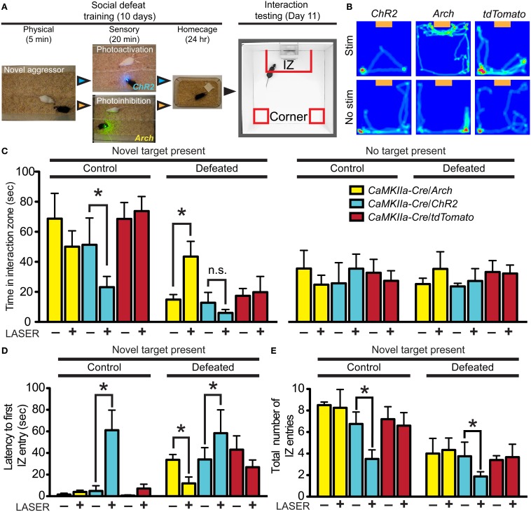
Divergent effects of vmPFC terminal photostimulation and photoinhibition in the DRN. (A) CaMKIIa-Cre mice were injected in the vmPFC with Cre-dependent AAVs coding for the expression of ChR2-YFP, Arch-GFP or tdTomato. Cohorts of mice were exposed to 5 min of physical defeat followed by 20 min of sensory contact with either photoactivation by ChR2 (473 nm, 10 mW, 25 Hz, 10 ms pulse width) or photoinhibition by Arch (561 nm, 10 mW) of vmPFC terminals via implanted fiber optic targeting the DRN. Mice were then placed in homecages overnight. This was repeated with exposure to a novel aggressor each day. On day 11, mice were assessed for approach or avoidance in the social interaction test. IZ—interaction zone. (B) Heat maps depicting representative behavioral effects of photoactivating (ChR2) or photoinhibiting (Arch) vmPFC terminals during the sensory period of social defeat on interaction with a novel social target (orange box). Red and green areas depict areas where mice spent the most time. No effect was observed in mice injected with sham tdTomato virus. (C) In defeated mice, photosilencing of vmPFC terminals prevented a decrease in social interaction (Two-Way ANOVA, Fisher post-hoc, *p = 0.050) while photoactivation did not decrease interaction times significantly from ChR2-expressing mice that did not receive laser stimulation. Undefeated control mice whose vmPFC terminals in the DRN were stimulated displayed a significant decrease in social interaction compared to unstimulated counterparts (Two-Way ANOVA, Fisher post-hoc, *p = 0.049). No effect of virus of photostimulation was observed when a novel social target was not present. (D) With a novel social target present, mice whose vmPFC terminals were photoactivated via ChR2 during sensory contact displayed significant increases in latencies to first enter the IZ in both control (Two-Way ANOVA, Fisher post-hoc, *p = 0.033) and defeated conditions (*p = 0.049). Defeated mice whose vmPFC terminals in the DRN were photosilenced via Arch during sensory contact displayed shorter latencies to first enter the IZ (*p = 0.048). (E) Total number of entries into the IZ was decreased in both control and defeated mice whose vmPFC terminals in the DRN were photoactivated during sensory contact (Two-Way ANOVA, Fisher post-hoc, control *p = 0.042, defeated *p = 0.049).

