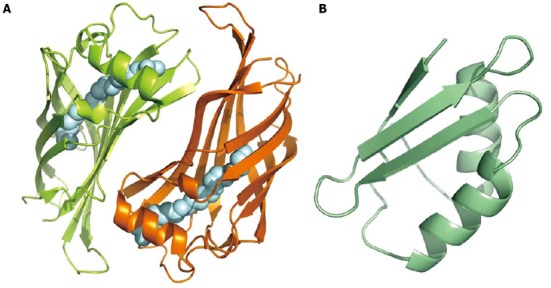
Figure 3.

Binding and transport proteins. A: Cartoon model of HP1286 lipocalin dimer. The two monomers, related by a two-fold axis, bind in the inner cavity a molecule of erucamide (silver spheres; PDB 3HPE); B: Nuclear magnetic resonance structure of apo-CopP, a copper binding regulatory protein of 66 amino acid residues (PDB 1YG0).
