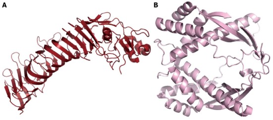
Figure 4.

Toxins. A: Cartoon model of the p55 domain of vacuolating toxin (VacA) (coordinates from PDB 2QV3). The structure is a predominantly right-handed parallel β-helix, and the domain mediates the binding of VacA to the host cell; B: Cartoon model of a truncated form of tumor-necrosis-factor α (TNFα) inducing protein, a virulence factor that enters gastric cells and stimulates both the production of TNFα and the nuclear factor kappa B pathway (coordinates PDB 2WCR).
