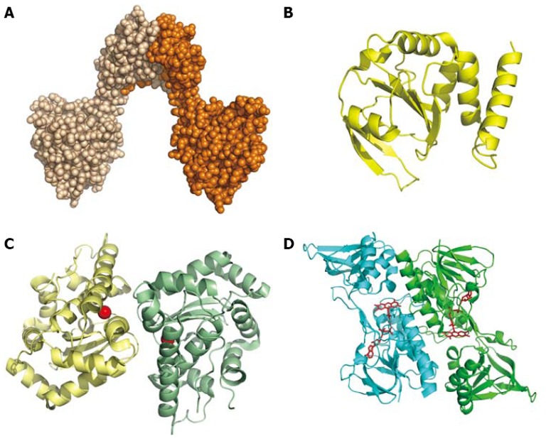
Figure 5.

Redox proteins. A: Space-filling model of the dimer of DsbG (HP0231; PDB 3TDG); B: Cartoon of DsbC (HP0377; Coordinates PDB 4FYC), an enzyme with a thioredoxin-like fold possibly involved in cytochrome c assembly; C: Cartoon of the dimeric Fe-superoxide dismutase (Coordinates PDB 3CEI). The iron ion is represented by a red sphere; D: Dimeric thioredoxin reductase (Coordinates PDB 3ISH). The FAD bound is shown as a ball-and-stick model.
