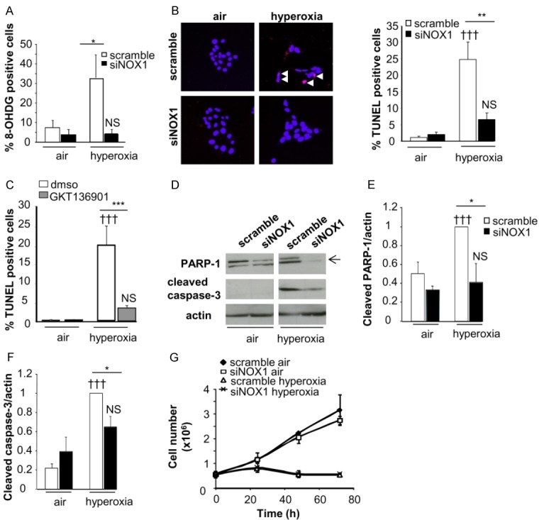
Figure 4.

Acute and stable NOX1 inhibition decrease hyperoxia-induced death of MLE12. Cell death was evaluated in control and NOX1-silenced MLE12 in air or hyperoxic condition. A: Transduced MLE12 exposed to air or hyperoxia for 72 h were stained with 8-hydroxy-2’-deoxyguanosine antibody (8-OHdG, red) and DAPI (blue) and the number of 8-OHdG-positive cells is expressed as percent of all nuclei (n>50 for each group, 3 independent experiments). B: Representative images of transduced MLE12 stained with TUNEL (red) and DAPI (blue) at 72 h of air or hyperoxia. White arrows indicate TUNEL-positive cells which appear in pink. The number of TUNEL-positive cells is expressed as percent of all nuclei (n>50 for each group, 3 independent experiments). P=NS, †††P<0.001 air versus hyperoxia; ***P<0.001, **P<0.01, *P<0.05 scramble-versus NOX1-silenced cells in hyperoxia. C: MLE12 were treated with DMSO, or GKT136901 (10 m) and exposed to hyperoxia for 72 h. ***P<0.001 cells treated with NOX inhibitor compared to cells exposed to DMSO in hyperoxia. P=NS, †††P<0.001 air versus hyperoxia. D-F: Protein lysate of transduced MLE12 were blotted for cleaved caspase-3 and PARP-1 and quantified by densitometry (right panel; n=3). β-actin was used to control equal loading. P=NS, †††P<0.001 air versus hyperoxia; *P<0.05 scramble-versus NOX1-silenced cells in hyperoxia. G: Cell growth was measured by using sulforhodamine B for different times. Absorbance was measured at 490 nm and cell number was determined. The relationship between cell number (protein content per well) and absorbance is linear from 0 to 5.106 cells. No difference was observed between scramble-versus NOX1-silenced cells in hyperoxia.
