
Figure 3.
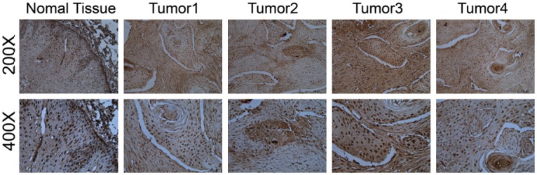
Representative IHC photos of TP53INP1 expression in normal tissue (200× and 400×) and ESCC tumors. TP53INP1 staining was mainly localized within the nucleus of cells in the form of yellow brown granules. It’s obvious that the positive staining intensity of ESCC is much more stronger than that of normal tissue.