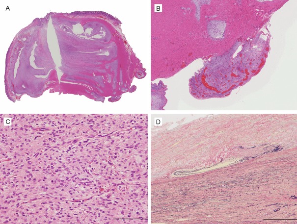
Figure 4.
Histological appearance of the tumor. A. The tumor shows a multinodular plexiform growth pattern. B. Fibromyxoid nodules of tumor extend into serosal surface. C. The tumor contains spindle-shaped bland tumor cells in a fibromyxoid stroma, which is rich in small caliber blood vessels. D. Although the tumor disrupted the vessel wall, there was no evidence of invasion of the intravascular lumen.

