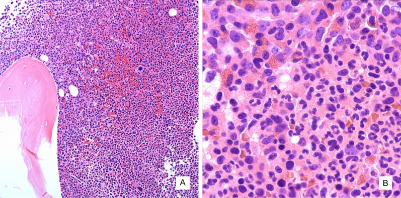
Figure 1.

Histomorphology of bone marrow biopsy. The marrow is hypercellular with increased myeloid to erythroid ratio and atypical megakaryocytes (A, hematoxylin and eosin stain, 100x). Eosinophils including immature eosinophilic myelocytes are focally increased (B, hematoxylin and eosin stain, 400x).
