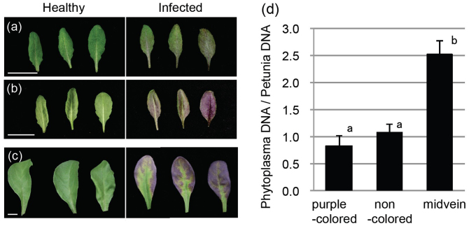
Figure 1. Purple top symptom and phytoplasma distribution.
Typical samples of OY-W-infected leaves exhibiting purple coloration symptom in Arabidopsis wild-type (a, b) and Petunia Vakara Blue (c). The leaves were healthy (each three leaves in left sides) and infected with OY-W phytoplasma (each three leaves in right sides). (a) shows upper surface of rosette leaves of Arabidopsis, and (b) shows lower surface. Bars = 1 cm. (d) Phytoplasma accumulation in Petunia leaves exhibiting purple coloration symptoms. Petunia leaves exhibiting symptoms were divided into three tissues; midveins, purple-colored leaf margins (purple-colored), and other non-colored leaf margins (non-colored). OY-W populations were estimated by real-time PCR for OY-W tufB gene. Each bar represents the average of three biological replicates (±SE). P. hybrida glyceraldehyde-3-phosphate dehydrogenase gene (PhGAPDH) was used for normalization. Different alphabets indicate significant differences between them (P < 0.05).

