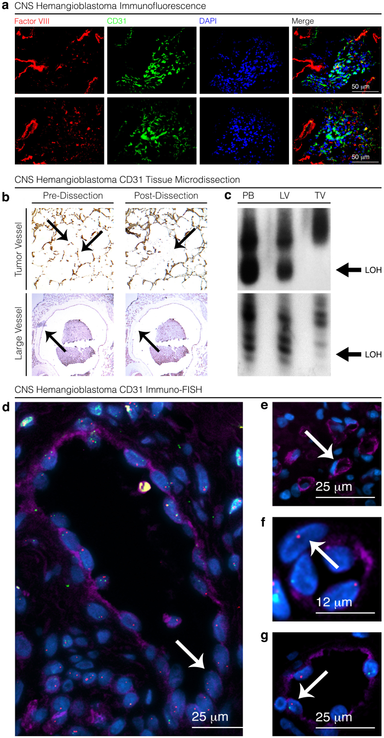
Figure 2. Analysis of endothelial cell origin in VHL-associated CNS hemangioblastoma.
Immunofluorescence staining reveals an absence of Factor VIII expression (red) in small (10–50 mm) CD31 expressing (green) vascular structures and minimal overlap in tumor-associated vessels (a). Micro-dissected vascular structures (arrows) within the tumor (TV), identified by CD31 staining, demonstrate marked allelic imbalance (loss of heterozygosity) (arrows) after amplification with D3S1038 and D3S1110 VHL-flanking chromosome 3 primers in comparison to large tumor-feeding vessels (LV) and blood samples (b,c). FISH analysis of CD31 staining endothelial cells (purple) confirming two copies of chromosome 3 (green) or VHL (red) (arrows) in most large vessel (100+ μm) endothelial cell nuclei (d) in comparison to loss of one copy of VHL (arrows) in nuclei of endothelial cells forming small (10–50 μm) vascular structures (e,f,g). Blots are cropped to enhance clarity and conciseness of presentation.

