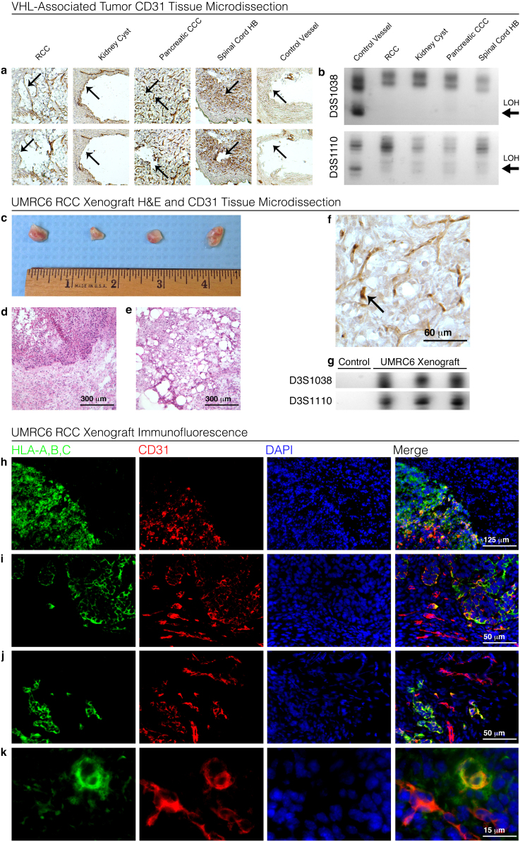
Figure 3. Evaluation of subcutaneously implanted UMRC6 murine xenografts for intratumoral vasculogenesis.
CD31 staining identifying vascular structures (arrows) within RCC, kidney cyst, pancreatic clear cell carcinoma, spinal hemangioblastoma, and non-tumor control vessel (a; upper row). CD31 staining vascular structures were micro-dissected and extracted (arrows) (a; lower row) and demonstrate LOH after amplification with D3S1038 and D3S1110 VHL-flanking chromosome 3 primers in comparison to non-tumor control vessel (b). Surgically extracted subcutaneous UMRC6 (renal cell carcinoma cell-line with loss of VHL) murine xenografts were assessed for intratumoral vasculogenesis (c). H&E staining of xenografts demonstrates sharp demarcation between human xenograft tumor cells and murine connective tissue (d), as well as characteristic VHL-associated tumor phenotype with the presence of clear-cells and extensive vascular tissue within the xenograft (e). Small vascular structures within the xenograft, identified through CD31 staining, were micro-dissected (f). Micro-dissected vessels demonstrate presence of human genomic (DNA) marker after amplification with D3S1038 and D3S1110, as compared to control murine muscle tissue that did not amplify (g). HLA immunofluorescence (green) clearly demarcated UMRC6 xenograft tumor cells from murine connective tissue cells (h). Co-localization by immunofluorescence for human-specific HLA (MHC class I A, B, and C) (green) and non-specific CD31 (red) confirmed the presence of human endothelial cells emerging within xenografts adjacent to invading murine (non-human) vascular tissue (i,j,k). Blots are cropped to enhance clarity and conciseness of presentation.

