Fig. 1.
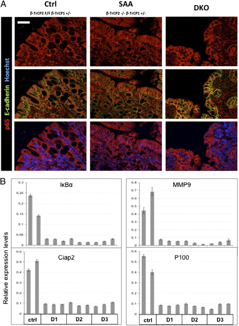
β-TrCP double KO in the gut results in NF-κB inhibition. (A) Immunofluorescence of colons from mice with the indicated genotypes. p65 (red), an NF-κB protein, is present in epithelial cell cytoplasm of control mice, with no NF-κB stimulus, and in the nuclei of inflamed mice (SAA) that have residual β-TrCP activity and normal, unstable IκB. In spite of the severe inflammation in the DKO mice, p65 is cytoplasmic, probably because of stabilized IκB. E-cadherin (green)-negative immune cells infiltrating the epithelial layer signify the inflammation in DKO colons. Blue color indicates Hoechst, a nuclear marker. (Scale bar: 50 μm.) (B) NF-κB target genes expression analysis in enterocytes from the indicated mice. Unlike control mice that show basal NF-κB target gene expression, in DKO mice, NF-κB transcriptional activity is totally inhibited starting at day 1 (IκBα; P = 0.16563; MMP9, P < 0.0001; Ciap2, P < 0.0001; P100, P < 0.0001; P values calculated by unpaired two-tailed t test, controls vs. all DKO samples regardless of the time of harvest).

