Fig. 2.
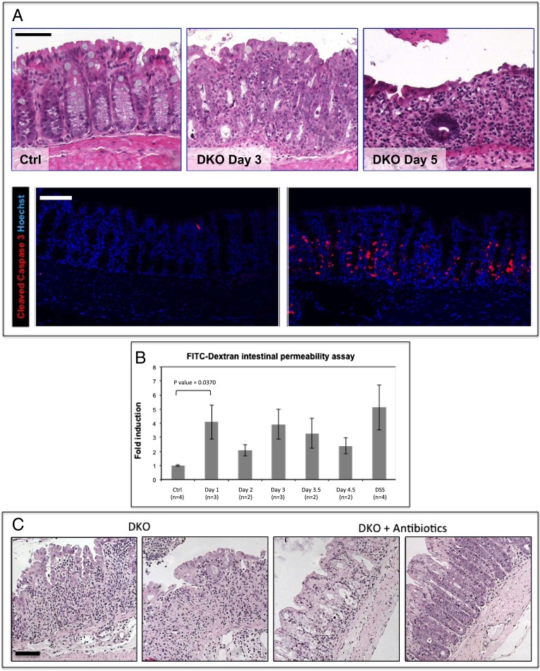
Barrier disruption and severe colitis in β-TrCP DKO intestines. (A) (Upper) H&E staining of colon sections of control mouse 5 d after tamoxifen injection (n = 21; Left), DKO mouse 3 d after injection (n = 19; Middle; tissue is abnormal with high numbers of mitotic and apoptotic cells and small immune cell clusters), and DKO mouse 5 d after injection (n = 34; Right). The epithelial layer is extremely thin, and the majority of cells are immune cells infiltrating the mucosa, which is hardly functional at this stage. (Lower) Cleaved caspase 3, an apoptotic marker (red) is abundant in day 3 DKO colons. Hoechst, a nuclear marker, is shown in blue. (Scale bars: 100 μm.) (B) Relative levels of FITC-dextran in the blood 6 h after FITC-dextran force-feeding of the indicated mice (Ctrl, negative control; β-TrCP1+/−, β-TrCP2f/f, no Cre). DSS is positive control. DSS-treated mice received 4% daily dosage for 5 d. DKO mice at day 1 show high intestinal permeability that is similar to the DSS-treated positive control (P = 0.0370 for day 1 DKO mice vs. controls, unpaired t test). (C) H&E staining of representative colons from DKO mice at day 5 (n = 3; Left) and DKO mice at day 5 treated for 15 d with antibiotic at day 5 (n = 3; Right). A significant alleviation of the pathologic process following antibiotic treatment; in contrast to DKO colons, in which hardly any epithelial cells are left at day 5, in the antibiotic-treated DKO colons, the epithelial layer is still present and is functional, although abnormal. (Scale bar: 100 μm.)

