Fig. 4.
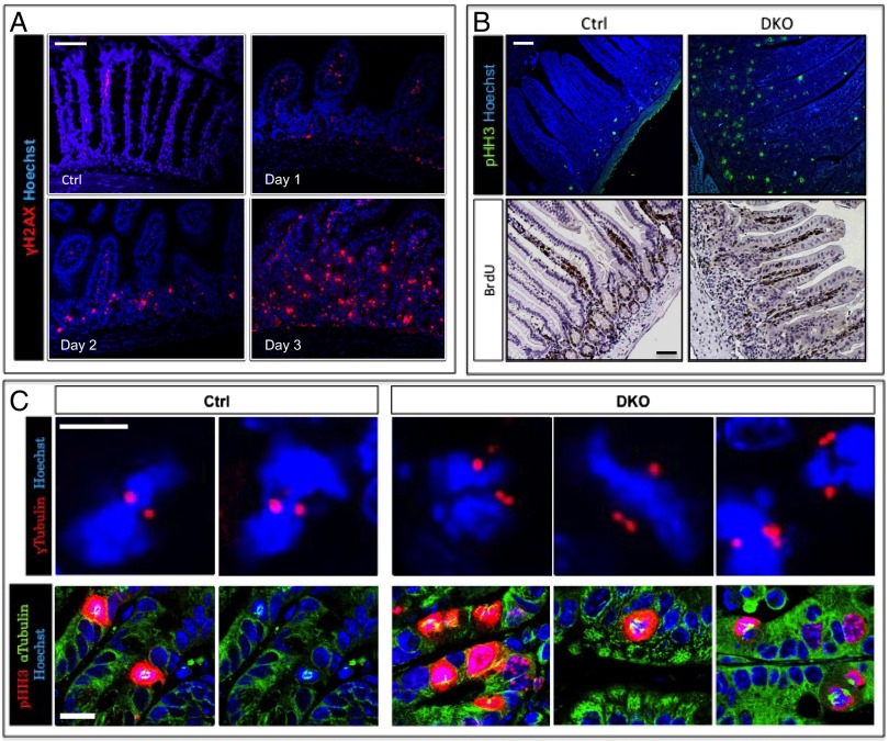
DNA damage is an early event following β-TrCP deletion. (A) Enterocytes positive for γ-H2AX, a DNA damage marker (red), are present in DKO intestines starting at day 1, and their numbers increase in later days. Hoechst, a nuclear marker, is shown in blue. (Scale bar: 100 μm.) (B) (Upper) High numbers of pHH3 (mitotic marker, green)-positive cells in DKO intestines compare with control as seen by immunofluorescence. Blue represents Hoechst, a nuclear marker. (Lower) IHC shows similar quantities of BrdU-positive cells (S-phase marker) in control and DKO intestines. The ratio between mitotic cells (i.e., pHH3-positive) and cells in S phase (i.e., BrdU-positive) is extremely high, indicating prolonged mitosis rather than high proliferation rate. (Scale bar: 100 μm.) (C) (Upper) Centrosome staining (γ-tubulin, red) pointing to more than two centrosomes per cell in DKO intestines (n = 3). No cells with more than two centrosomes were found in control mice (n = 3). Nuclei are stained by Hoechst (blue). (Scale bar: 10 μm.) (Lower) Mitotic cells (pHH3, red) with abnormal spindles (α-tubulin, green) in DKO mice. No abnormal spindles were found in control mice. Nuclei are stained by Hoechst (blue). (Scale bar: 20 μm.)

