
Fig. 5.
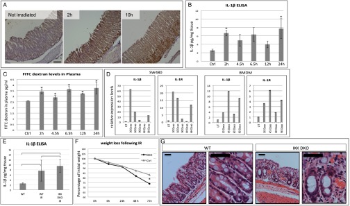
DNA damage can induce IL-1β up-regulation in an NF-κB–independent manner. (A) IL-1β IHC demonstrates IL-1β up-regulation in WT mice in response to DNA damage induced by 4 Gy γ-irradiation. The times indicated are number of hours after irradiation. (Scale bar: 100 μm.) (B) IL-1β ELISA on intestinal supernatants of WT irradiated mice. As soon as 2 h after 6 Gy γ-irradiation, IL-1β secretion is elevated more than twofold compared with nontreated mice (n = 3 for all groups; 2 h, P = 0.0109; 4.5 h, P = 0.1166; 6.5 h, P = 0.1361; 12 h, P = 0.2500; 24 h, P = 0.0915; P values calculated by unpaired two-tailed t test). (C) FITC-dextran permeability assay on WT irradiated mice. Gut permeability is increased following 6 Gy γ-irradiation (n = 3 in all groups; 2 h, P = 0.0017; 4.5 h, P = 0.0291, 6.5 h, P = 0.0019; 12 h, P = 0.0002; 24 h, P = 0.0212; P values calculated by unpaired two-tailed t test). (D) qPCR results provide evidence to IL-1β mRNA up-regulation shortly after doxorubicin treatment (5 μg/mL) in SW480 intestinal cell line (Left) and primary bone marrow-derived macrophages (Right). DOXO was given in a 15-min treatment at 1 μg/mL Time points are in minutes unless otherwise indicated. (E) IL-1β ELISA on intestinal supernatants of untreated WT, γ-irradiated WT, and γ-irradiated IKKα/IKKβ DKO mice. γ-Irradiation at 9 Gy induced similar elevation in IL-1β secretion in intestines with intact NF-κB or lacking any NF-κB activity, indicating an NF-κB–independent IL-1β secretion. P = 0.2027, WT vs. gamma-irradiated WT mice (WT IR). P = 0.0174, WT vs. IKK DKO IR. P = 0.4424, WT IR vs. IKK DKO IR. P values were calculated by unpaired two-tailed t test and are indicated in the chart. (F) Weight loss of WT and IKK DKO mice following 9 Gy γ-Irradiation. IKK DKO mice lost more weight than their WT littermates. P = 0.0031 at the day of euthanasia. (G) H&E staining of WT and IKK DKO large intestines 3 d after 9 Gy γ-irradiation. The pathology in IKK DKO mice is more severe, with increased inflammatory infiltrates. (Right) Higher-magnification image of each genotype. (Scale bar: 50 μm.)