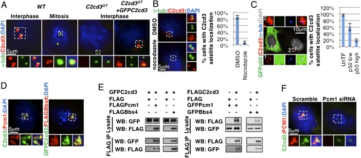
Fig. 1.
C2cd3 is localized to centriolar satellites. (A) In interphase, C2cd3 is present in punctae around centrosomes, whereas in mitosis, C2cd3 is localized to two punctae at each spindle pole. The lack of staining in C2cd3GT mutant cells indicates the specificity of the C2cd3 antibody, and GFP–C2cd3 exhibits similar localization to endogenous C2cd3. (B) The centriolar satellite C2cd3 is dispersed upon nocodazole treatment, revealing its centriolar localization. DMSO-treated cells served as a control. (C) The overexpression of p50/Dynamitin disrupts the centriolar satellite localization of C2cd3. (D) C2cd3 is colocalized with Pcm1 and Bbs4. (E) Overexpressed C2cd3 coimmunoprecipitates with Pcm1 and Bbs4. (F) C2cd3 is dispersed from centriolar satellites in cells treated with a Pcm1 siRNA. The lack of Pcm1 staining indicates the effectiveness of the knockdown. Both Pcm1 and C2cd3 are localized to centriolar satellites in the control cells treated with a scramble siRNA. Centrosomes and spindle poles are labeled with γ-tubulin. The nuclei are visualized with DAPI. For quantitative analyses, SD is indicated. n = 3 independent experiments. IP, immunoprecipitation; WB, Western blot.

