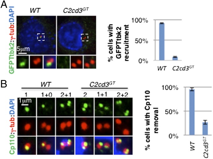
Fig. 3.
Ttbk2 recruitment and Cp110 removal are compromised in the absence of C2cd3. (A) GFP–Ttbk2 is localized to the centriole in wild-type cells but not in C2cd3GT mutant cells. (B) Cp110 is removed from the mother centriole in wild-type cells but not in C2cd3GT mutant cells. Note that Cp110 staining is dynamic through the cell cycle. Before centrosome duplication, Cp110 was observed as one dot in wild-type cells, representing the daughter centriole (“1”). In contrast, two dots were observed in C2cd3GT mutant cells, representing both the mother and daughter centrioles (“2”). After centrosome duplication, only one centrosome is positive for Cp110 (“1+0”) in wild-type cells, but both centrosomes are positive for Cp110 (“1+1”) in C2cd3GT mutant cells. In the G2 phase, the procentrioles are capped with Cp110 such that Cp110 appears as two dots in the centrosome containing the daughter centriole, and one dot representing the procentriole in the centrosome containing the mother centriole (“2+1”) in wild-type cells. In contrast, C2cd3 mutant cells exhibit two Cp110 dots in each centrosome in the G2 phase (“2+2”). The quantitative analysis includes interphase cells. The centrosomes are labeled with γ-tubulin, and the nuclei are visualized with DAPI. For quantitative analyses, SD is indicated. n = 3 independent experiments.

