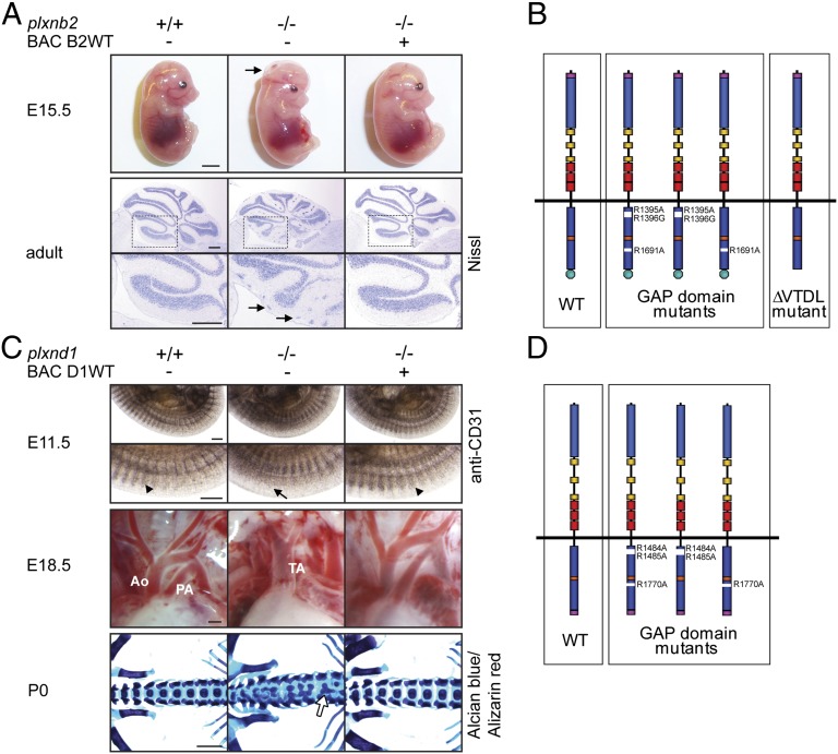
Fig. 1.
Generation of BAC transgenic mice expressing triple-myc-tagged versions of Plexin-B2 and Plexin-D1. (A) (Upper) Pictures of E15.5 embryos; arrow points to the open cephalic neural tube (exencephaly). (Lower) Nissl-stained adult cerebella; boxed areas are magnified below, arrows point to clusters of ectopic granule cells at the cerebellar surface. (Scale bars, 3 mm in Upper, 300 µm in Lower.) (B) Schematic illustration of the allelic series of plxnb2 BAC transgenes. Purple, triple-myc-tag; blue, Sema domain; yellow, PSI domains; red, IPT/TIG domains; dark blue, split GAP domain; orange, Rnd/RhoD/Rac1 binding site; green, PDZ domain interaction motif. (C) (Top) WhoIe-mount immunohistochemistry on E11.5 embryos using an anti-CD31 antibody. Arrowheads point to intersomitic vessels, and arrow points to disorganized intersomitic vessels. (Middle) Pictures of the cardiac outflow tract of E18.5 embryos. Ao, Aorta; PA, pulmonary artery; TA, truncus arteriosus. (Bottom) Alcian blue/Alizarin red staining of P0 skeletons. Arrow points to fusion of vertebral bodies. (Scale bars, 300 µm in Top and Middle, 1 mm in Bottom.) (D) Schematic illustration of the allelic series of plxnd1 BAC transgenes. Purple, triple-myc-tag; blue, Sema domain; yellow, PSI domains; red, IPT/TIG domains; dark blue, split GAP domain; orange, Rnd/RhoD/Rac1 binding site.

