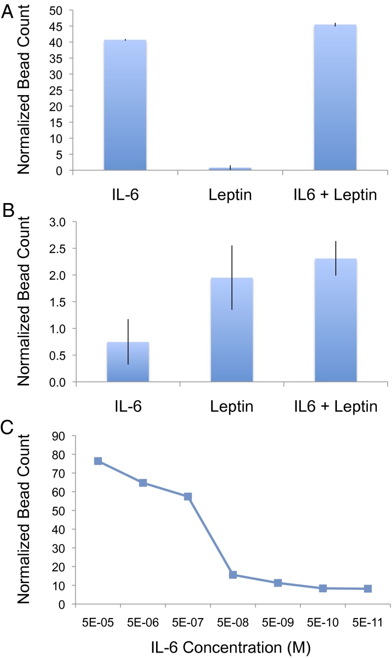
Fig. 3.
IL-6 and leptin microfluidic immunoassays; 50% human sera was spiked with recombinant IL-6 (5 nM), leptin (600 nM), or IL-6 and leptin mixed together. Each sample was analyzed using both anti–IL-6 (A) and anti-leptin (B) chips to demonstrate specificity of our microfluidic sandwich immunoassay approach. Titrating concentrations of IL-6 were also analyzed using anti–IL-6 chips under the same conditions to evaluate dynamic range and sensitivity (C). Detection of the bound cytokine of interest was performed using anti–IL-6– or anti-leptin–conjugated beads. Bound beads were optically quantified, and normalized bead counts were calculated by dividing by the number of beads observed to nonspecifically be bound in a control channel (no antigen, 50% human serum) performed in parallel. A dynamic range of at least six orders or magnitude and a limit of detection of 50 pM was observed.

