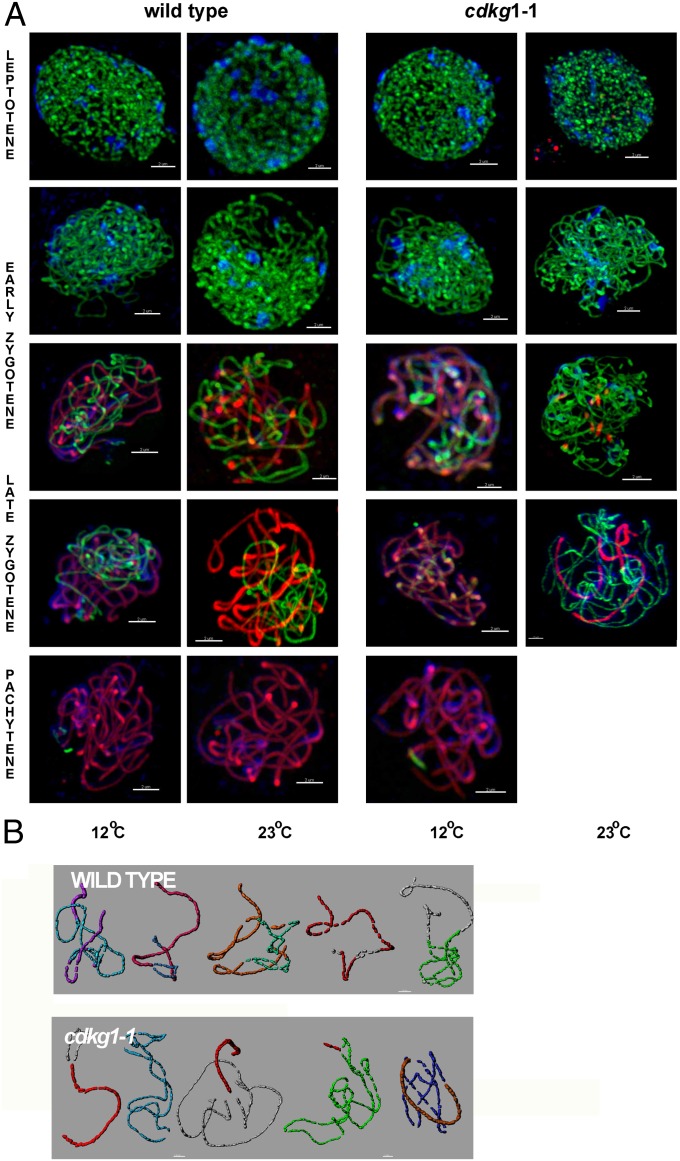
Fig. 3.
Synaptonemal complex formation in WT and cdkg1–1 mutant meiocyte nuclei, isolated from plants grown at 12 or 23 °C. (A) Nuclei at different meiotic stages immunostained with ASY1 (green) and ZYP1 (red) with DAPI counterstain (blue). (Scale bar, 2 μm.) (B) Three-dimensional reconstruction of individual bivalents from a pachytene WT nucleus (Upper) and pachytene-like mutant nucleus (Lower) at 23 °C. The nuclei were processed using Imaris, and each bivalent pair was isolated and false-colored. Quantification of ZYP1 loading is given in Fig. S3.

