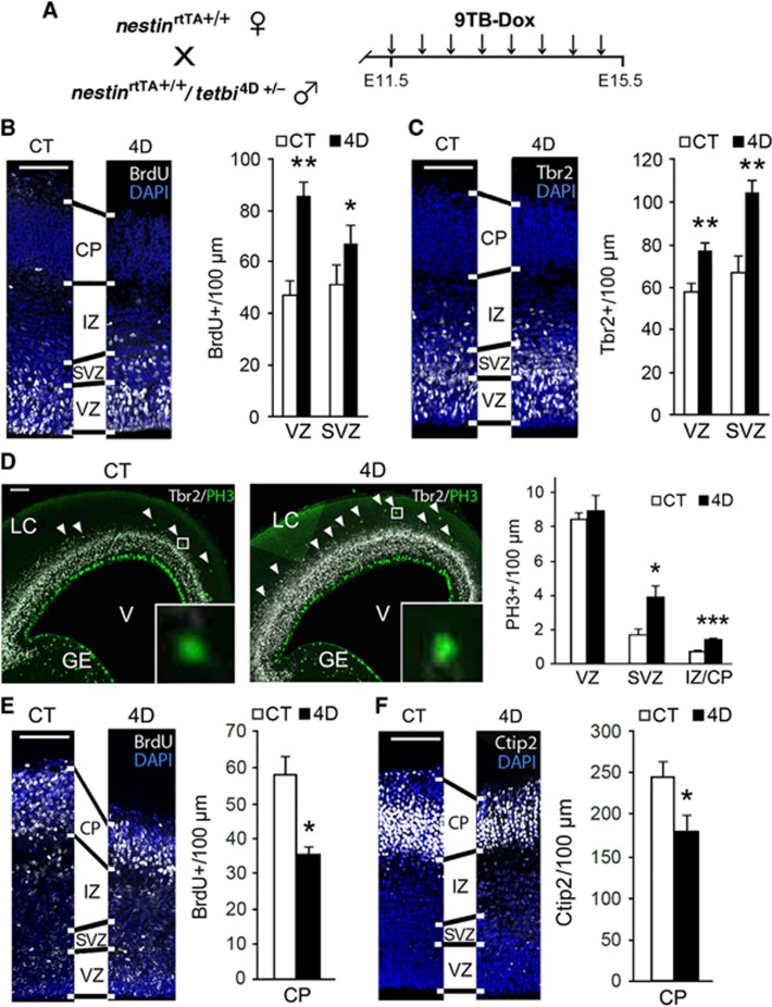
Figure 2.
BPs expand at the expense of neurogenesis in 4D transgenic mouse embryos. (A) Experimental approach used to study the effect of 4D overexpression after continous administration of 9TB-Dox from E11.5 to E15.5, followed by sacrifice. (B–F) Fluorescence pictures and quantifications of cells labelled for various markers, as indicated in each panel, in cortices of E15.5 CT and 4D littermate embryos treated with 9TB-dox starting at E11.5. BrdU was administered either 5 h prior to sacrifice (B) or at E12.5 (E). Shown in each panel are coronal sections taken from an equivalent cortical region of CT and 4D littermates as identified by stereology. Note the change in thickness of cortical layers in each image pair (lines between panels). Scale bars, 100 μm. Data are mean±s.e.m; *P<0.05; **P<0.01, ***P<0.001; n>3 pairs of embryos from different litters. See also Supplementary Figure S1.

