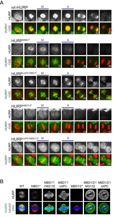
Figure 5. Microtubule-binding stabilizes HURP during mitosis.
A. HeLa cells were transduced with lentiviruses expressing mCherryhistone H2B and GFP-tagged HURP, HURPMBD1*, HURPΔAPC/MBD1/2*, HURPMBD1/2*, or HURPΔAPC/MBD1/2* (MBD1/2*: mutation of microtubule-binding motifs; ΔAPC: mutation of all degrons). Progression through mitosis was monitored by live cell imaging. The upper panels show the levels of the GFP-tagged HURP variant, whereas lower panels display GFP-HURP (green), mCherry-histone H2B (red), and the time post nuclear envelope breakdown. M: first frame with metaphase chromosome alignment; A: first frame with sister chromatid separation. B. HeLa cells were transfected with FLAGHURP, FLAGHURPMBD1* or FLAGHURPMBD1/2*; where indicated, APC/C-degrons were mutated (FLAGHURPΔAPC/MBD1*; FLAGHURPΔAPC/MBD1/2*) or cells were treated with MG132. Cells were analyzed by immunofluorescence microscopy. Upper panel: HURP-proteins; lower panel: HURP (green); tubulin (red); DNA (blue). See also Figure S4.

