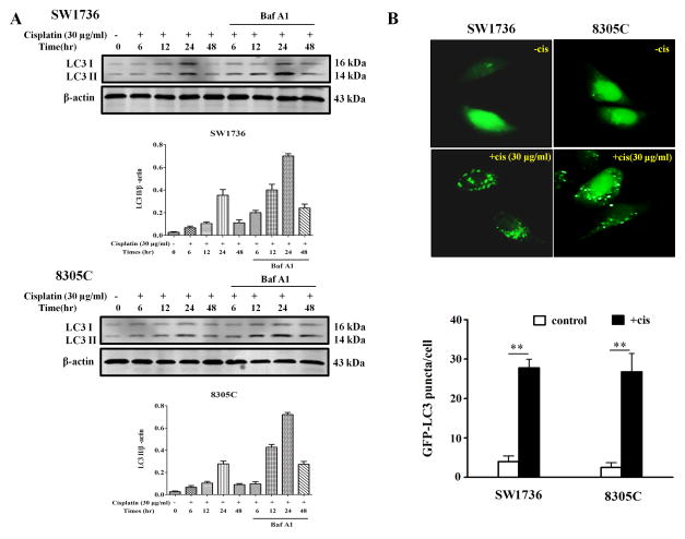
Figure 2. Cisplatin induces autophagy in anaplastic thyroid cancer (ATC) cells.
(A) SW1736 and 8305C cells were treated with 30 μg/ml of cisplatin for 6h, 12h, 24 h and 48h in the absence or presence of 10 nM of bafilomycin A1. At the end of treatment, cell lysates were prepared, resolved by SDS–polyacrylamide gel electrophoresis and subjected to western blot analysis using anti-LC3 or β-actin antibodies, respectively. β-actin was used as a loading control. Data shown are the representative of three identical experiments. (B) SW1736 and 8305C cells were transfected with a GFP-LC3 plasmid, followed by treatment with the indicated concentration of cisplatin for 24 h. At the end of treatment, the cells were inspected under a fluorescence microscope. Quantitation of the GFP-LC3 puncta was performed by counting 20 cells for each sample, and average numbers of puncta per cell were shown. The bars are the mean±s.d. of triplicate determinations; results shown are the representative of three identical experiments. **P<0.01, t-test, cisplatin versus vehicle.

