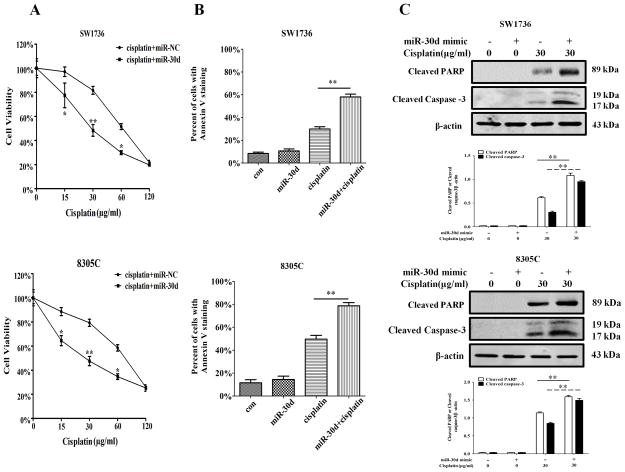
Figure 5. MiR-30d mimic increases sensitivity of ATC cells to cisplatin.
SW1736 and 8305C cells transfected with a mimic of miR-30d (100 nM) or a control RNA (100 nM) were treated with the indicated concentrations of cisplatin for 48 h. At the end of treatment, (A) cell viability was measured by 3-(4,5-dimethylthiazol-2-yl)-2,5-diphenyltetrazolium bromide assay. Each point represents mean±s.d. of triplicate determinations; results shown are the representative of three identical experiments. *P<0.05 and **P<0.01; (B, C) apoptosis was determined by flow cytometric analysis of Annexin V staining (B) and by western blot analysis of cleaved PARP and cleaved caspase-3 (C). β-actin was used as a loading control. Data shown are the representative of three identical experiments. **P<0.01, t-test.

