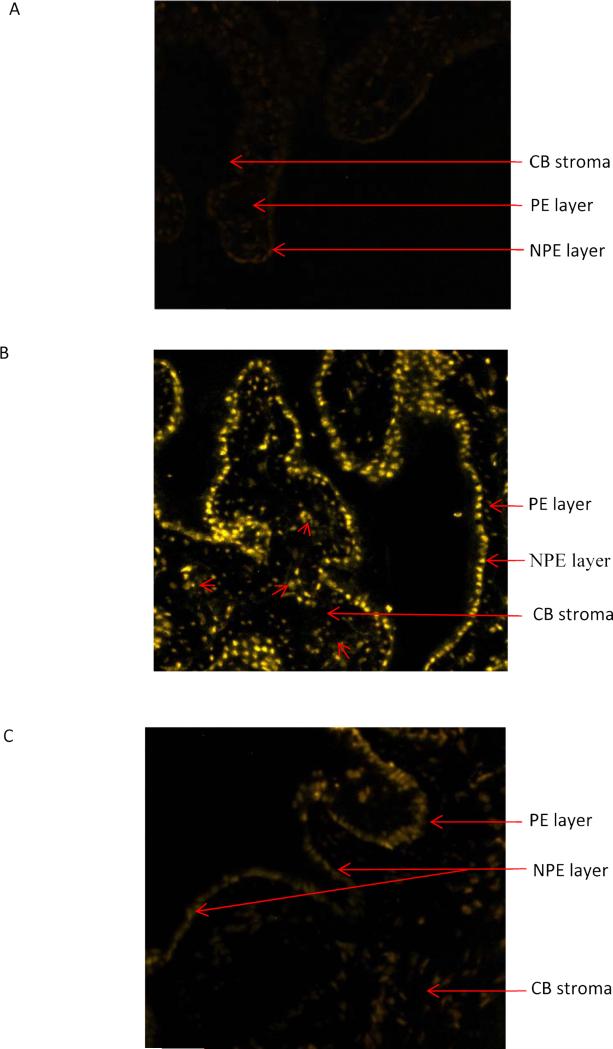
Figure 2.
Propidium iodide (PI) uptake by ciliary processes in the intact porcine eye. The aqueous humor compartment was perfused with control medium for 90 min. After this equilibration period, the eyes were perfused for 40 min with control or calcium-free medium that contained propidium iodide (~25 μM). Panel A shows propidium iodide detected by confocal microscopy in 7 micron thick sections obtained from eyes perfused with control medium. Panel B shows propidium iodide detected in eyes perfused with calcium-free medium plus 1 mM EGTA. In eyes perfused with calcium-free medium propidium iodide is detectable mainly in the NPE cell layer. In some regions, propidium iodide is also detected in the PE (arrows) and ciliary process stroma (arrowheads). Panel C shows the results obtained from eyes perfused with calcium-free medium containing 18α-glycyrrhetinic acid (100 μM) (AGA).

