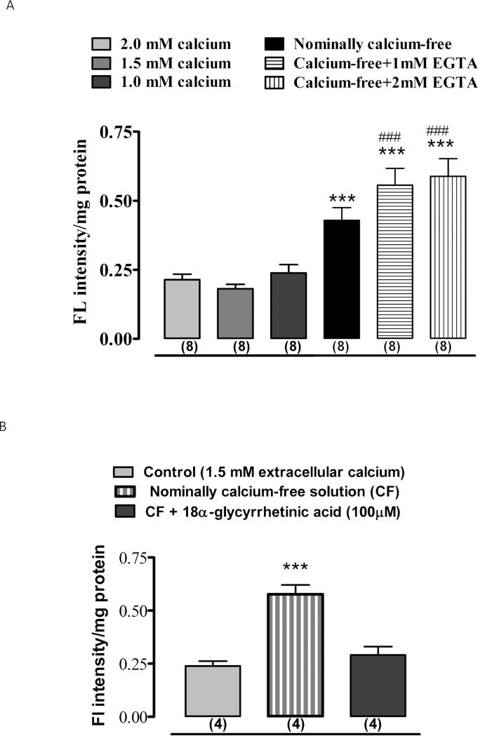
Figure 3.
The sensitivity of propidium iodide uptake by cultured NPE to extracellular calcium and 18α-glycyrrhetinic acid. Cells were exposed to 25 μM propidium iodide for 30 min in control (calcium-containing) medium, nominally calcium-free medium or in calcium-free medium containing EGTA (1-2 mM) added to chelate trace amounts of calcium. Quantitative data on propidium iodide fluorescence under these conditions is shown in panel A. Panel B shows the results obtained from cells incubated in nominally calcium-free medium containing 18α-glycyrrhetinic acid (100 M).The values are the mean ± SE of results from 4-8 independent experiments. *** indicates a significant difference (p<0.001) from control and ### indicates a significant difference (p<0.001) from nominally calcium-free condition.

