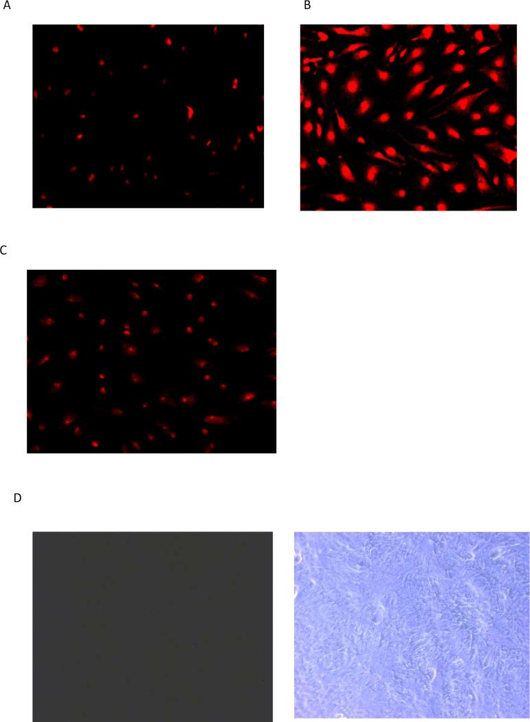
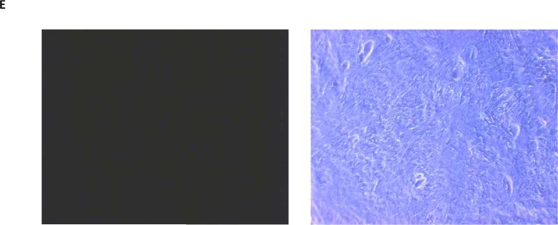
Figure 4.
Propidium iodide (PI) uptake by cultured NPE shown in low power micrographs. Panel A shows micrographs of cells exposed to propidium iodide (25 μM) for 30 min in control medium. Panel B shows micrographs of cells exposed to propidium iodide in nominally calcium-free medium. Panel C shows the results in calcium-free medium containing 18α-glycyrrhetinic acid (100 μM). Propidium iodide, which is associated with cell nuclei, was most abundant in the calcium-free group. In a separate experiment cells were exposed to fluorescein dextran (4000 μM) for 30 min in control or calcium-free medium then examined by fluorescence and phase contract microscopy. Cells from the control group and calcium-free group are shown in Panels D and E respectively with the phase contrast images on the right.


