
Figs. 1a - 1d.
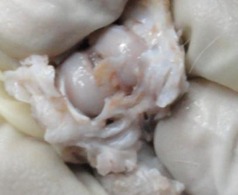
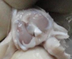
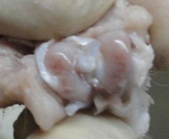
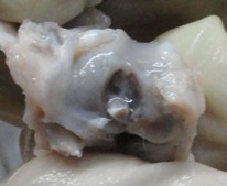
Figures 1a and 1b – gross photographs of femoral condyles in the mesenchymal stem cell (MSC)-treated group a) at 16 weeks post-operatively, showing a reduction in lesion severity and b) at 20 weeks post-operatively, showing further reduction in lesion severity. Figures 1c and 1d – gross photographs of femoral condyles in the control group at c) 16 and d) 20 weeks post-operatively, showing characteristics of osteoarthritis (OA) becoming more evident between time-points.