
Figure 6.
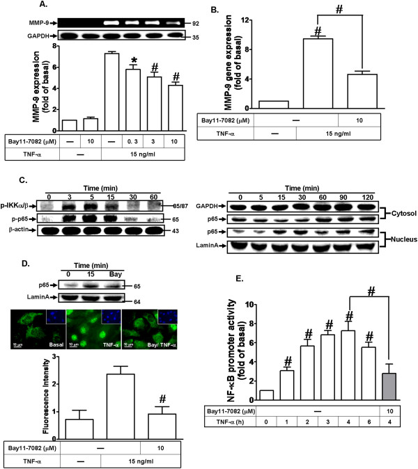
NF-κB is required for TNF-α-induced MMP-9 expression. (A) Cells were pretreated with Bay11-7082 for 1 h and then incubated with TNF-α (15 ng/ml) for 24 h. MMP-9 expression was determined as described in Figure 1. (B) Cells were pretreated with Bay11-7082 (10 μM) for 1 h and then incubated with TNF-α (15 ng/ml) for 6 h. The isolated RNA samples were analyzed for the levels of MMP-9 mRNA by real-time PCR. (C) Cells were incubated with TNF-α (15 ng/ml) for the indicated time intervals. The cell lysates were analyzed by Western blot using an anti-phospho-IKKα/β, anti-phospho-p65, or anti-β-actin antibody (left part). The cytosolic and nuclear fractions were analyzed by Western blot using an anti-NF-κB (p65), anti-Lamin A, and anti-GAPDH antibody (right part). (D) Cells were pretreated without or with Bay11-7082 (10 μM) for 1 h and then stimulated with TNF-α (15 ng/ml) for 15 min. The nuclear fraction was analyzed by Western blot using an anti-NF-κB (p65) and anti-Lamin A antibody. The translocation of p65 NF-κB was also observed by immunofluorescence staining (middle part) and the histogram of p65 translocation (lower part). (E) Cells were transiently transfected with NF-κB-Luc construct and then incubated with TNF-α (15 ng/ml) for the indicated time intervals in the absence or presence of Bay11-7082 (10 μM). The cell lysates were collected and determined the luciferase activity. Data are expressed as mean±SEM of three independent experiments. #P < 0.01, as compare to the cells treated with TNF-α alone. *P < 0.05, as compare to the cells treated with TNF-α (A,B,E) or vehicle alone (E).