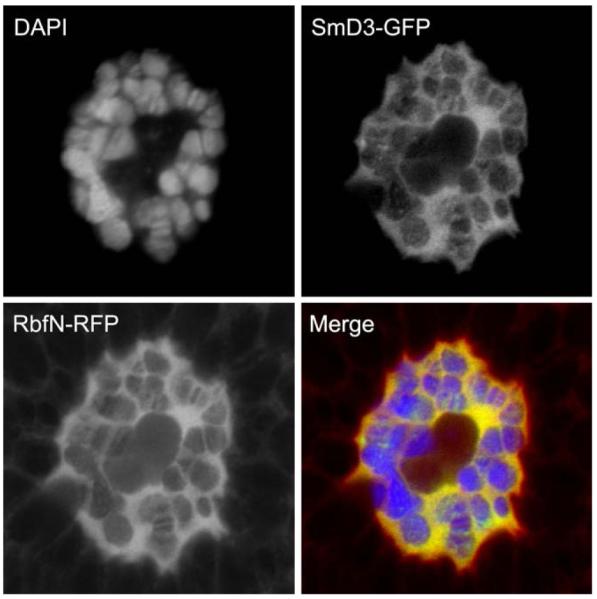
Fig. 1.

The Drosophila retinoblastoma tumor suppressor colocalizes with the spliceosomal component SmD3 in salivary gland nuclei. The amino-terminal domain of Drosophila Rbf fused to RFP (RbfN-RFP) colocalizes with a GFP-tagged SmD3 in larval salivary gland cells. The DNA stain DAPI was used to show the bands of the characteristic polytene chromosomes. Confocal imaging shows extensive colocalization of SmD3-GFP and Rbf1N-RFP in the nucleoplasm and along the chromsomes. Salivary glands were isolated from mature larvae and fixed with 4% formaldehyde. Confocal images were obtained using a Zeiss LSM510 Meta.
