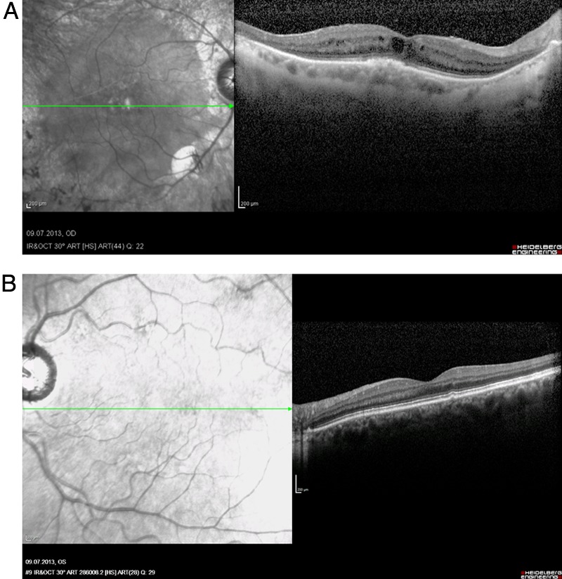
Figure 5.

Spectral-domain optical coherence tomography of the macula (right eye, 2013). (A) Right eye: cystoid oedema of the macula and atrophy of the inner retinal layers in the peripheral macula. (B) Left eye: single, parafoveal drusen formation, otherwise normal retinal structure.
