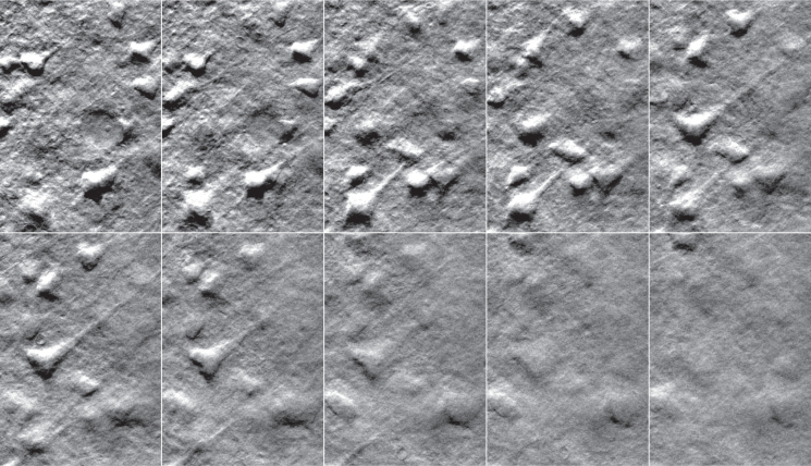
Fig. 2.
OBM phase-gradient images of mouse pyramidal neurons demonstrating optical sectioning. The frames are selected from a z-stack obtained by scanning the tunable lens ( Media 1 (3MB, AVI) ). The images were taken at equal intervals along a span of 42 μm (displayed here at intervals of 4.7 μm) at increasing depths from top-left to bottom-right. The field of view for each image is 67×96 μm. The total exposure time per frame was 20 ms (10 ms per active LED).

