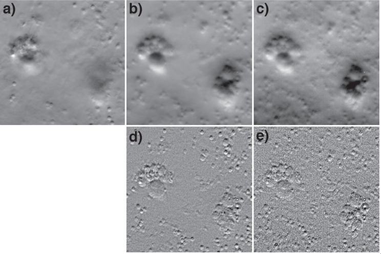
Fig. 3.
EDOF images of polystyrene (2 μm) and glass (3–10 μm) beads embedded in agarose gel. Panel a is a standard OBM phase-gradient image. Panels b,c are EDOF phase-gradient images acquired in single exposures by scanning the focal plane over ranges 25 μm and 100 μm respectively. Panels d,e are the same EDOF images, but deblurred according to Eq. (18). The field of view for all panels is 63×63 μm.

