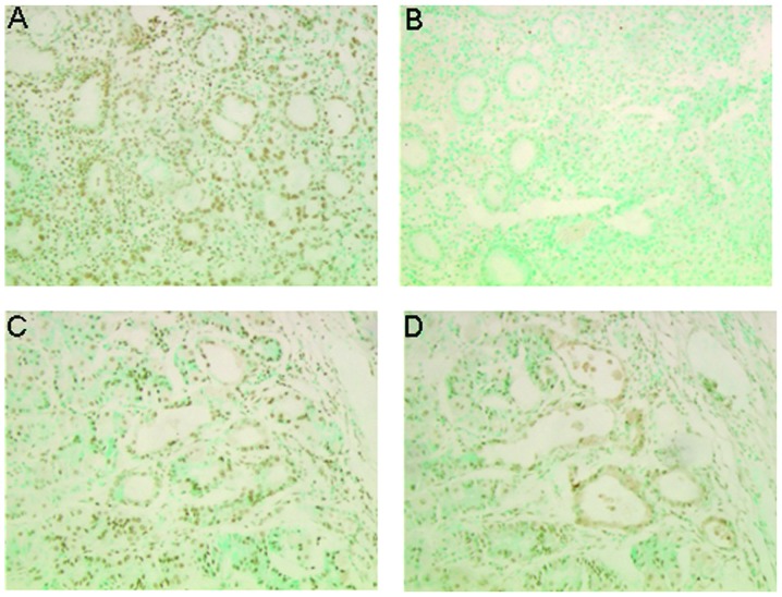
Figure 1.
Analysis of the apoptotic activity of the PAT-SC1 antibody in vivo. (A and B) A pre-treatment biopsy and (C and D) a post-treatment tumour sample of a stomach carcinoma patient were investigated for PAT-SC1-induced apoptosis using the Klenow FragEL DNA fragmentation kit (Oncogene, Boston, MA, USA). (A and C) In the positive controls, all cell nuclei are stained due to treatment with an endonuclease. (B and D) In the images (right panels) only the nuclei of apoptotic stomach tumour cells are stained. (B) The pre-treatment biopsy shows no apoptotic activity. (D) In the post-treatment tumour sample an increase in apoptotic tumour cells after PAT-SC1 treatment is noted.

