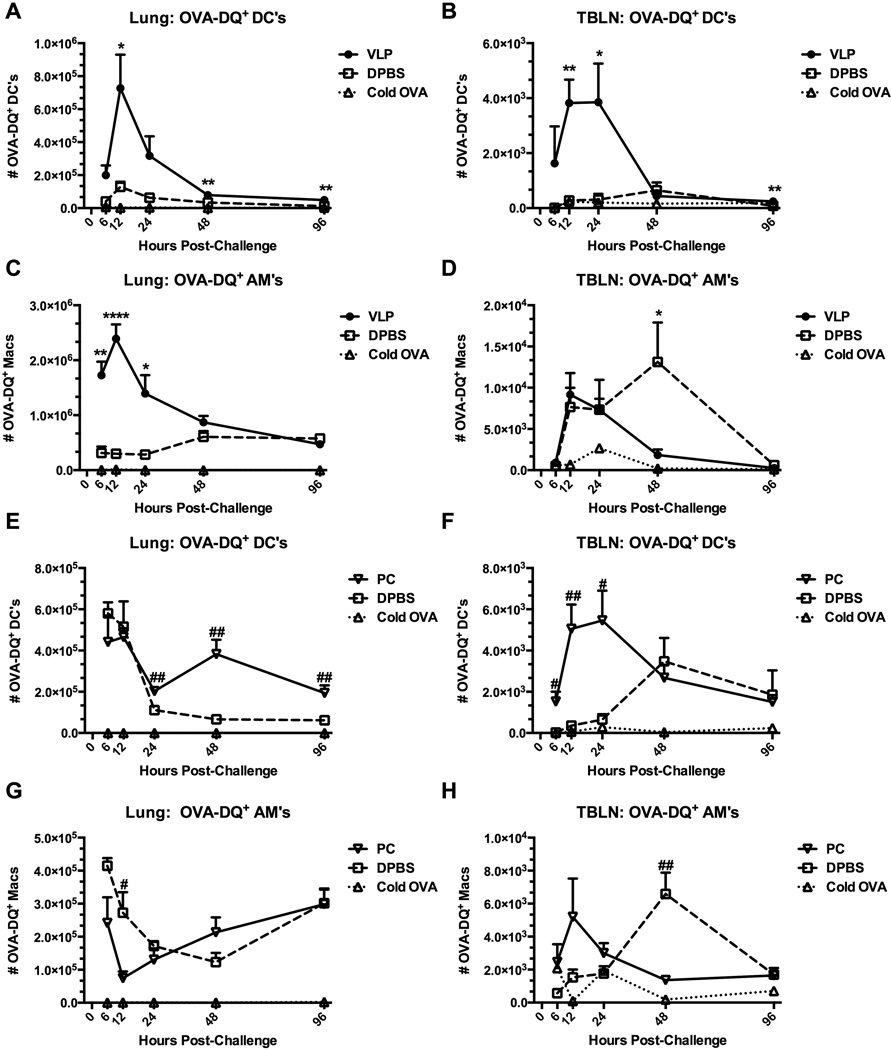
Figure 3.
Exposure of the lungs to VLPs, or infection with Pneumocystis causes enhanced antigen processing in response to an unrelated challenge. Mice were exposed to VLPs or vehicle (A-D) (C57BL/6), or infected with Pneumocystis (PC) (E-H) (BALB/c). Recovered mice were then i.n. challenged with 100 µg Ovalbumin-DQ, or unlabeled regular “cold” OVA as a staining control, and sacrificed at designated timepoints. OVA-DQ+ DCs (A&B, E&F) and AMs (C&D, G&H) in whole lungs (left panel) and TBLNs (right panel) were quantified by FACS and total hemocytometer cell counts. DCs and AMs were identified as CD11c+ and Siglec-F (DCs) or Siglec-F+ (AMs) from plots previously gated on forward and side scatter. Total cells were additionally quantified by total hemocytometer counts. Data are shown as mean + SEM of n=5. Experiments of similar design were independently performed at least twice. Significance as compared to the DPBS control group (unpaired t-test) is indicated by “#” for PC-infected mice and “*” for VLP-exposed mice.

