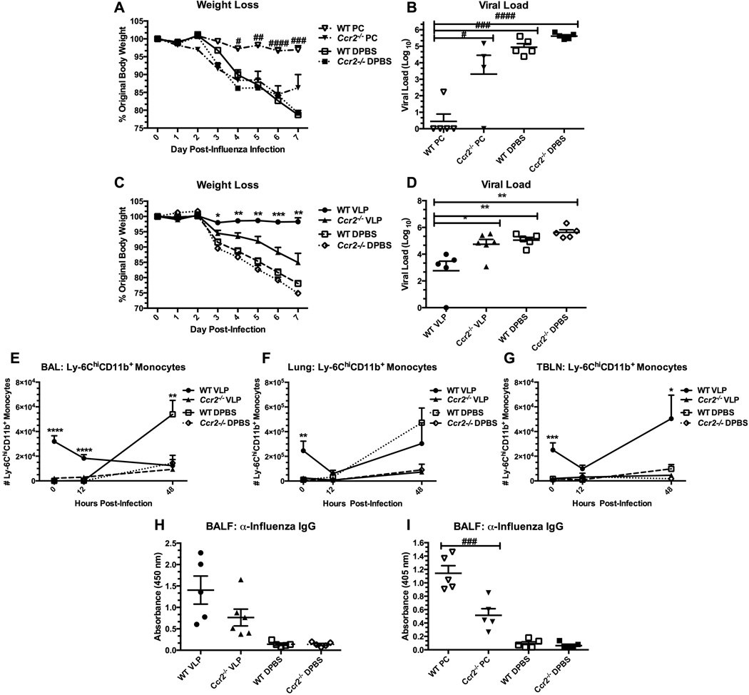
Figure 6.
Ccr2−/− mice are not protected from influenza virus via prior exposure to VLPs, or Pneumocystis infection. C57BL/6 (WT) or Ccr2−/− mice were infected with Pneumocystis (PC), or exposed to VLPs, or vehicle. Recovered mice were then challenged with 1.5 × 10³ pfu PR8. Mice were weighed daily post-influenza infection (A&C), and viral load from the left lung lobe was determined at day 7 post-infection (B&D). In a second study of similar design (VLPs only), groups of mice were killed at 0, 12 hours, and 48 hours post-infection and the multilobe of the lungs and whole TBLNs were collected and prepared for FACS. Cells isolated from BAL (E), lung homogenate (F), and TBLN (G) were stained for Ly-6C+CD11b+ monocytes. Granulocytes were gated from forward and side scatter plots and resultant CD11clo/int cells were gated and analyzed for their expression of Ly-6C and CD11b. For antibody determination, cell-free lavage fluid was analyzed by ELISA to determine the concentration of anti-influenza IgG (H&I). In figure 6H, BALF samples were diluted 1:2 prior to analysis samples in figure 6I were plated neat. Data are shown as mean + SEM of n=5 from One-way ANOVA analysis with a Bonferroni’s post-test. Significance as compared to the DPBS control group per strain is indicated by “#” for PC-infected mice and “*” for VLP-exposed mice. Experiments of similar design were independently performed at least twice.

