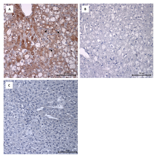
Figure 1.
Immunocytochemistry of RHDV antigen in the liver. A: Light micrograph of a liver paraffin section of an immunosuppressed, RHDV-infected young rabbit that died with RHD showing widespread positive immunodetection of the major RHDV antigen (brown staining). Heterophils are observed surrounding the damaged hepatocytes and inside sinusoids (arrows). The tissue section was labelled with an anti-VP60 mouse antiserum; haematoxylin counterstain; bar = 100 μm. B: A liver section of an immunosuppressed, RHDV-infected young rabbit that died with RHD and that was labeled with control mouse antiserum; haematoxylin counterstain, bar = 100 μm. C: A liver paraffin section of an infected young rabbit that survived the infection and was euthanized 7 days later. Signs of hepatocellular regeneration are visible as expressed by enlarged and often binucleated cells (without the formation of organized hepatic cords); some tissue spaces are occupied by small basophilic cells, with an oval shape, that are suggestive of hepatocyte precursors. The tissue section was labeled with an anti-VP60 mouse antiserum; haematoxylin counterstain, bar = 100 μm.

