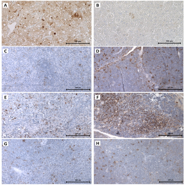
Figure 2.
Immunocytochemistry of apoptosis in the liver, spleen and thymus. Light micrographs of the liver, spleen and thymus paraffin sections where the immunodetection of cellular apoptosis was obtained by TUNEL; counterstaining with haematoxylin. In liver sections, bar = 100 μm; in spleen and thymus sections, bar = 200 μm. A: A liver paraffin section of an immunosuppressed, RHDV-infected young rabbit that died due to RHD. B: A liver paraffin section of an immunosuppressed, RHDV-infected young rabbit that died from RHD; control of the TUNEL assay. C: A spleen paraffin section of an immunosuppressed young rabbit euthanized 7 days after immunosuppression. D: A thymus paraffin section of an immunosuppressed young rabbit euthanized 7 days after immunosuppression. E: A spleen paraffin section of an immunosuppressed, RHDV-infected young rabbit that died from RHD. F: A thymus paraffin section of an immunosuppressed, RHDV-infected young rabbit that died from RHD. G: A spleen paraffin section of an infected young rabbit euthanized 7 days after RHDV infection. H: A thymus paraffin section of an infected young rabbit euthanized 7 days after RHDV infection.

