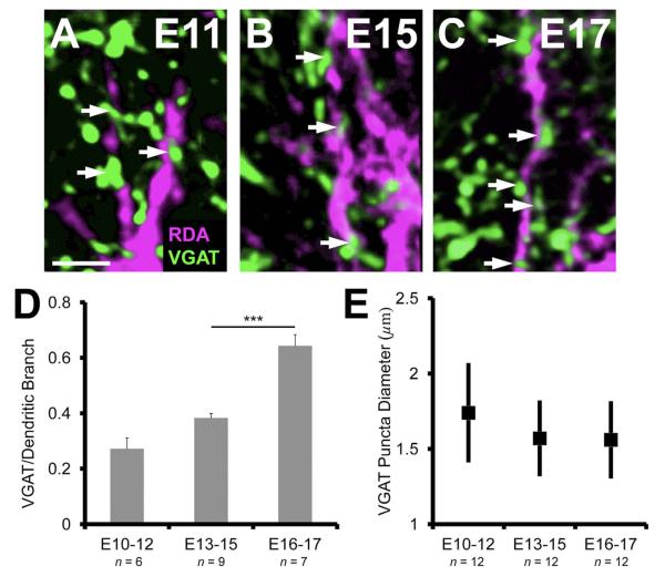
Figure 4.
VGAT apposition along NL dendrites increases at late embryonic ages. A–C: The progressive increase in the number of VGAT puncta (white arrows) per unit length (μm) of NL dendrite between E11 and E17. D: The increase from E10–12 (27.2 ± 3.9%) to E13–15 (38.3 ± 1.6%) is not significant (P = 0.053). The number of VGAT apposed to dendrites at E16–17 (64.3 ± 4.1%) is significantly greater (P < 0.0002) than previous ages, suggesting a key phase in inhibitory synapse development. E: The range in puncta size did not change over age (1.3–2.3 μm in diameter, ANOVA, P = 0.21). Scale bars = 10 μm.

