Figure 5.

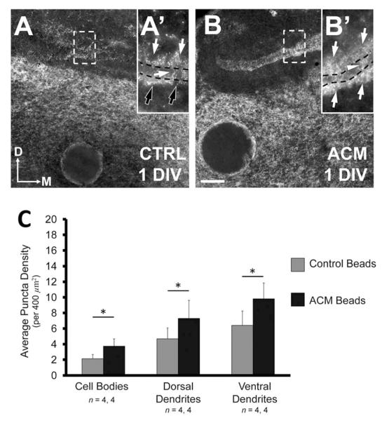
Astrocyte-secreted factors increase the density of VGAT in vitro. A: Rostral slices (see Materials and Methods) after +1 DIV with control medium exhibit a distribution of VGAT density similar to that observed in vivo at E13–15 with a greater density of inhibitory synapses in the ventral neuropil (white arrowheads) than in the dorsal neuropil (white arrows), and cell body region (black arrow). B: Rostral slices treated with ACM for +1 DIV exhibit an increase in VGAT density in the dorsal and ventral neuropil (white arrows). ACM does not increase the number of inhibitory terminals within the cell body region (black arrow). C: Caudal slices (see Materials and Methods) incubated for +1 DIV with control medium exhibit the same polarized distribution in the ventral neuropil (white arrowheads). D: The density of VGAT within the dorsal and ventral dendritic regions of caudal slices also increases after +1 DIV with ACM, but not on the cell bodies (black arrow). In both B,D, the increase is such that both dorsal and ventral dendritic regions exhibit similar inhibitory terminal density (white arrows). E: Change in density after +1 DIV with ACM compared to controls in each NL region. Slices treated with ACM had a significantly greater density in both the ventral (from 6.21 ± 0.43 puncta per 400 μm2 to 10.83 ± 0.88, P < 0.0002) and dorsal dendritic region (from 5.61 ± 0.35 to 10.48 ± 0.97, P < 0.0002) compared to control slices. The difference between the density in the cell body region of treated and control slice was not different (from 4.25 ± 0.45 to 6.19 ± 0.84, P = 0.10, Wilcoxon T). There was a significantly greater density of inhibitory terminals within the ventral and dorsal dendritic regions than observed in the cell body region in treated slices (P < 0.002 and P < 0.02, respectively). Scale bar = 20 μm.
