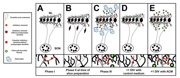
Figure 7.
Summary of interactions that take place during development and in organotypic slice experiments. A: The first phase of inhibitory synaptogenesis is the period between E10–12. SON axons are in contact with neuron in NL and inhibitory terminals (red circles) just begin to populate the ventral neuropil and cell body layer. B: We place brainstem slices into culture during phase 2, at E13, prior to the emergence of GFAP-positive astrocytes. While there has been an increase in the number of terminals within the ventral and dorsal neuropil, there remains a pronounced asymmetry with a greater density of VGAT puncta in the ventral neuropil (black arrows). SON axon terminals may not extend into the dorsal neuropil (dashed axon extension), alternatively, or dorsally extending terminals may not yet express VGAT. Terminals may not yet be associated with a postsynaptic dendrite (red circles traced in black). C: After phase three (E16–17), inhibitory terminals are evenly distributed throughout both the ventral and dorsal dendritic regions. At this age GABAergic release sites are closely aligned with their dendritic targets and the synapses function to depolarize compartments of NL dendritic arbors. The density of terminals remains significantly less within the cell body region than in the cell-free neuropil. This distribution may be due, in part, to astrocytes (blue patches) that surround NL and secrete factors (green circles) that interact with NL cell bodies and their dendrites. D: After +1 DIV, in the absence of any astrocyte-secreted factors, there is a small increase in the number of terminals along the ventral neuropil and the imbalance between dorsal and ventral dendrites is still apparent. The number of VGAT puncta along NL dendrites has not changed. E: The last panel represents what we hypothesize results from astrocyte-secreted factors in the developing NL. SON axons have branched into the dorsal dendrite region and new terminals can be found along NL dendrites. Evidence presented here suggest that factors contained with ACM from GFAP-positive astrocyte isolated from E16 brainstems are capable of mimicking the developmental increase and distribution of inhibitory inputs to NL observed during the last phase of inhibitory synaptogenesis.

