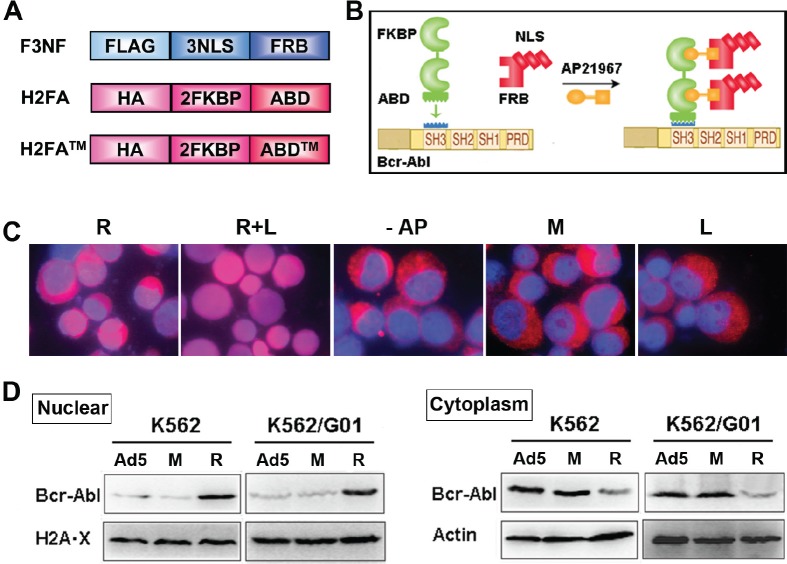
Figure 1. RNTS directs Bcr-Abl from cytoplasm into the nucleus.
(A) Schematic illustration of recombinant proteins F3NF, H2FA and H2FA™. (B) The schematic diagram of RNTS mediated transportation of Bcr-Abl into the nucleus. This figure was derived from modification of the AP21967 instruction picture of Clotech. (C) Nucleocytoplasmic transport was investigated in K562 cells by immunofluorescent assay. Images were merged for two colors: DNA(blue) and Bcr-Abl (red). R: RNTS, L: LMB, AP: AP21967, M: RNTS™, Ad5: Ad5 adenovirus vector. These abbreviations will be used in the rest of the figures. (D) Nuclear and cytoplasmic proteins were collected separately. The amount of Bcr-Abl in the nucleus and cytoplasm was quantified by western blot.

