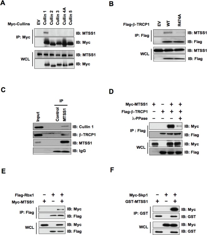
Figure 1. SCF complex containing β-TRCP1 and Cullin 1 interacts with MTSS1.
(A) Immunoblot (IB) analysis of whole cell lysates (WCL) and immunoprecipitates (IP) derived from 293T cells transfected with Myc-tagged Cullin constructs or empty vector (EV) as a negative control. (B) IB analysis of WCL and IP derived from 293T cells transfected with Flag—tagged wild-type or R474A mutant -TRCP1 constructs, or EV as indicated. (C) 293T cell extracts were immunoprecipitated with antibody against MTSS1, or control IgG and analyzed by IB analysis. (D) IB analysis of WCL and IP derived from 293T cells transfected with Myc-MTSS1 and Flag—β-TRCP1 constructs as indicated. Where indicated, cell lysates were pre-treated with λ-phosphatase before the IP procedure. (E) IB analysis of WCL and IP derived from 293T cells transfected with Myc-MTSS1 and Flag-Rbx1 constructs, as indicated. (F) IB analysis of WCL and IP derived from 293T cells transfected with GST-MTSS1 and Myc-Skp1 constructs, as indicated.

