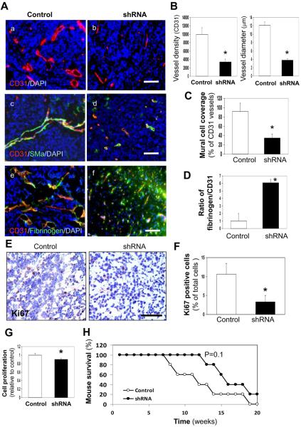
Figure 1. YKL-40 expression in GSDC-transplanted tumors is associated with vascular stability, mural cell coverage, angiogenesis, and tumor growth.
A. Representative immunofluorescent images of control and YKL-40 shRNA GSDC brain tumor sections from SCID/Beige mice depicted single staining of CD31 (red) (a, b) and double staining of CD31 (red) with either SMa (green) (c, d) or fibrinogen (green) (e, f). DAPI (blue) was used to stain the nuclei. B. Quantification of CD31 vessel density and vessel diameter from A (a, b) as described in the Methods. The latter was an average of individual luminal diameters. C. Quantification of percent mural cell coverage of CD31 vessels from A (c, d). The data were derived from the ratio of SMa density to CD31 density. D. Quantification of the ratio of fibrinogen vs. CD31 for vessel leakiness from A (e, f), in which the ratio of fibrinogen density to CD31 density in the control tumors was set as 1 unit. E. Representative control and YKL-40 shRNA GSDC tumor staining images of the proliferation marker Ki67. F. Percentage of Ki67 positive cells with brown nuclear staining was quantified. G. Cell proliferation in culture using MTS assay. N=12. H. Kaplan-Myer Survival curve of SCID/Beige mice bearing control or YKL-40 shRNA tumors. N=5. *P≤0.05 compared to corresponding controls. Bars: 100 μm.

