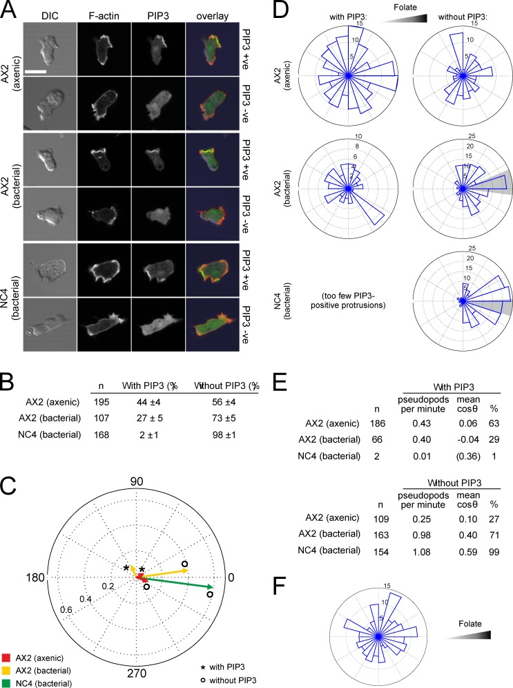
Figure 2.
PIP3–labeled protrusions do not orient up folate gradients. (A) AX2 and NC4 cells transfected with the F-actin reporter Lifeact-mRFP and the PIP3 reporter PH-CRAC-GFP were examined by confocal microscopy, and the presence or absence of a PIP3 patch in each cell was determined during random migration. Bar, 10 µm. (B) Numbers of pseudopods with and without PIP3 patches during random migration. Percentages are the means and standard deviations from three experiments. (C and D) Mean direction of protrusions during folate chemotaxis. (C) The displacements from the migration vectors of all pseudopods were measured, and the mean is shown in a circular plot. If all protrusions were extended in the same direction, a vector with length 1 would result. PIP3-containing pseudopods from NC4 were too rare to give a reliable direction. (D) Rose plots of the directions of all protrusions with and without PIP3. The shaded area indicates the 95% confidence interval of the mean angle. (E) Frequency and direction of pseudopods in C and D. (F) Rose plot of the direction of PIP3 patches that form macropinosomes during folate chemotaxis.

